E-ISSN:2583-9152

Research Article

Organic, Inorganic & Biomaterials

Journal of Condensed Matter

2024 Volume 2 Number 1 Jan. - June
Publisherwww.thecmrs.in

Study of perylene Di-imide as a Binder of a Human Telomeric Tetraplex DNA by Molecular Docking Simulation

Mishra V1*, Tiwari R2
DOI:https://doi.org/10.61343/jcm.v2i01.49

1* Vandana Mishra, Department of Physics, DDU Gorakhpur University, Gorakhpur, U P, India.

2 Rakesh Kumar Tiwari, Department of Physics, DDU Gorakhpur University, Gorakhpur, U P, India.

Human telomeres are consisting of d(TTAGGG) repeats involved in the formation of tetraplex DNA structures. Ligands stabilizing these tetraplex DNA structures are potential inhibitors of the cancer cell-associated enzyme telomerase. In human cells, telomerase adds multiple copies of the 5’-GGTTAG-3’ motif to the end of the G-strand of the telomere and in the majority of tumour cells it results over-expressed. Several structural studies have revealed a diversity of topologies for telomeric tetraplexes, which are sensitive to the nature of the cations present, to the flanking sequences, and probably also to concentration, as confirmed by the different conformations deposited in the Protein Data Bank (PDB). The existence of   different polymorphism   in the DNA quadruplex and the absence of a uniquely precise binding site give rise to a better understanding of mechanism by verifying with docking methodology approach. To target this, we have selected six different experimental models of the human telomeric sequence d[AG3(T2AG3)3] with the ligands (the   perylene di-imide). We simulated out molecular docking of binding of perylene di-imide to a selected G-quadruplex using dock 6.9 to examine the or loop binding mode of perylene di-imide.

The simulation provides the two highest rank docking poses of perylene di-imide i.e. with external strand and groove binding mode; the role different ligand binding is described as external striking on the terminal guanine tetrad and the groove binding. which may be further considered for the study of therapeutic properties of ligand.

Keywords: DNA Duplex, Docking, telomeric quadruplex and molecular interaction

Corresponding Author How to Cite this Article To Browse
Vandana Mishra, , Department of Physics, DDU Gorakhpur University, Gorakhpur, U P, India.
Email:
Mishra V, Tiwari R, Study of perylene Di-imide as a Binder of a Human Telomeric Tetraplex DNA by Molecular Docking Simulation. J.Con.Ma. 2024;2(1):20-23.
Available From

Manuscript Received Review Round 1 Review Round 2 Review Round 3 Accepted
2024-02-06 2024-04-02 2024-04-22
Conflict of Interest Funding Ethical Approval Plagiarism X-checker Note

© 2024by Mishra V, Tiwari Rand Published by Condensed Matter Research Society. This is an Open Access article licensed under a Creative Commons Attribution 4.0 International License https://creativecommons.org/licenses/by/4.0/ unported [CC BY 4.0].

Introduction

G-tetraplex DNA is a high concentration structure of G-Prosperity nucleic acid sequence. They are held together by a hoogesteen type of hydrogen bond and stabilized by monovalent cations [1]. These structures are found at the ends of eukaryotic chromosomes and-MYC, C-kit, KRAS and BCL-2 Such as are found in affected areas called promoter region [2]. G-tetraplex structures may also play a role in the translation process, which are potentially present in messenger RNA. Stable tetraplex has been shown to effectively prevent telomerase activation, which is often overexpressed in human cancers [3].

In solution four-Repetitive human telomere sequences, d[AG3(T2AG3)3], first of NMR-Characteristics of based structure1993 Was made in [4]. This sequence is an intramolecular-forms G-quadruplex in which three stacked G-Tetrads are involved with anti Syn-Syn around each tetrad glycosidic structure. The adenine bases of loop Through stacking interactions with G-tetrads let's talk, a so-called basket structure(pdb143D) [5].

In 2002, By Professor Needle's group in a K+ the different sequence of the same sequence in the compound crystal [6]. G- quadruplex structure was observed: all four strands are parallel, Connecting TTA loop double chain-are reversers, and all guanine based Let's adopt anti- glycosidic structure (pdb1KF1), Later studies physiological K+ for several human telomere sequences in solution conditions Indicated the presence of a mixture of G- quadruplex forms [7].

Other than this, NMR analysis revealed that the telomeric sequence contains other intramolecular conformations, which are defined as hybrids. Small ligands can promote the formation of quadruplex structures from telomeric DNA, can inhibit telomerase enzyme and cause telomere ends in cancer cells, may destabilize end-capping [8]. Based on this, Design of new ligands as potential anticancer agents.  Several studies have been conducted to synthesize and evaluate G-quadruplex ligands as potential anti-cancer agent [9].

In this perspective, we come across a website on which structure of many drugs were made. from here we downloaded the structure of perylene di-imide [PDIs] and got it docking simulation with human telomeric sequence d[AG3(T2AG3].

Perylene Di-imide (PDIs)

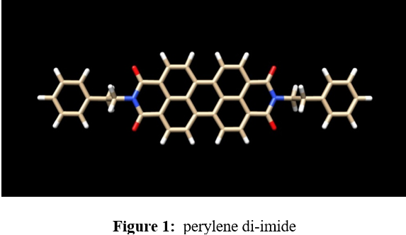
The structure of perylene di-imide is shown in figure 1.

manoj.png

Perylene di-imide is also known as N-N di-(2phenylethyl) perylene carbonic acid, amide. Molecular formula is C40H26N2O4 [PUBCHEM]. PDIs are highly electron deficient conjugated structure that can exist stably in ambient environments. PDIs also targets binding of terminal telomeric G-quadruplex inhibiting telomerase [10].

Methodology

Receptor preparation

Downloaded structures from the Protein Data Bank (PDB) including 1KF1, 4wo3, and NMR models 143d, 2l7v, 2mb3, 2mgn, and 2o3m related to the telomeric sequence d[AG3(T2AG3)3] underwent pre-treatment. Co-crystallized water molecules and counter ions were removed from these structures. Additionally, any sequences containing hybrid models were noted to have head and tail caps, each consisting of a different number of additional nucleotides.

Ligand preparation

The perylene di-imide depicted in figure 1 was utilized in the current study. The ligand structure was obtained by downloading it from the PUBCHEM website. Subsequently, the structure with the lowest energy conformation was selected. This chosen conformation was then prepared for further docking studies, maximizing torsion, using the dock 6.9 tool.

Docking Conditions 

In the final step, the docking approaches employed involved screening all generated configurations. Configurations within 3 kcal mol-1 above the global minimum energy complex were subjected


to full minimization using the same force field and environment described for the target optimization procedure. Analysis of the results incorporated consideration of thermodynamic estimates derived from state equations, including free energy, enthalpy, and entropy of complex formation computed at 300 K.

Three-dimensional figures were generated using CHIMERA graphics, as depicted below. All calculations were executed on a Linux cluster.

Results and Discussion

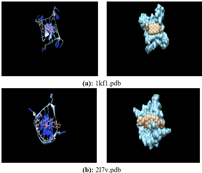
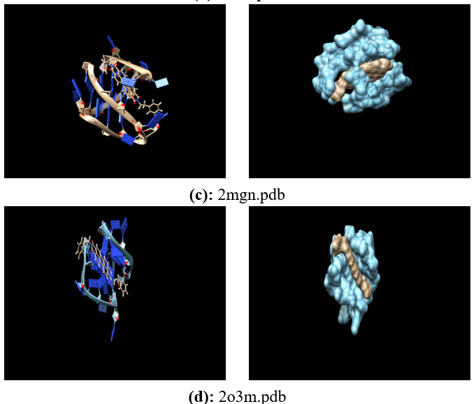
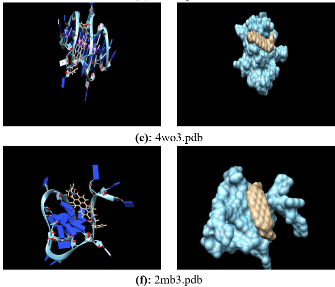
Considering this observation, we decided to include it in the docking analysis with the widely used method Dock 6.9. The human telomeric repeat sequence, d[AG3(T2AG3)3], has been experimentally characterized in various conformations. To account for conformational variability, we included six PDB models (1kf1, 2L7v, 2mgn, 2o3m, 4wo3, 143d, 2mb3) representing both X-ray and NMR structures, specifically on G-tetrad structures be focused shown in figure 2.

26-1.png

26-2.png

26-3.png

Figure 2: Six PDB models representing both X-ray and NMR structures.

Evaluation of the most stable conformations for each model was conducted after the pretreatment, which included structural optimization through energy minimization. Below is included an image showing various G-Tetraplex complexes with perylene di-imide. In each displayed image, the view on the left presents the standard perspective provided by the CHIMERA software, while the view on the right presents a surface view also generated by CHIMERA. The depicted images depict complex structures, where the sky-blue colour represents the receptor and the brown colour represents the ligand, specifically perylene di-imide.

Conclusion

In conclusion, we modelled the G-tetraplex fold, using experimentally described


conformations of the human telomeric repeat sequence d[AG3(T2AG3)3] based on the six G-tetrads available in protein data bank (PDB), The importance of considering diverse forms has been underlined. The chemical compound chosen as our ligand is PDIs.

Our primary motivation for conducting this study was to investigate whether molecular docking simulations using Docked 6.9 could recapitulate the binding mode observed in the crystal structure of the human telomeric G-quadruplex with PDIs.

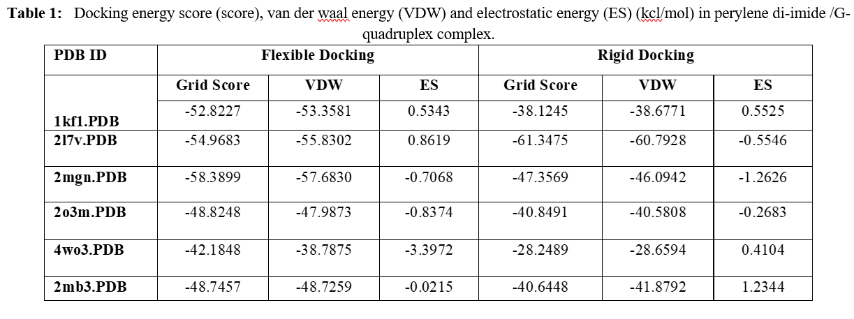
Our molecular docking simulations generated different binding modes, both flexible and rigid in nature. The corresponding docking poses are illustrated in the figures, and the docking results are summarized in table 1. The presented docking energy scores represent the cumulative values of Vander Waals (VDW) and electrostatic (ES) terms.

26-4.png

References

1. Alcaro S, Costa G, Distinto S, Moraca F, Ortuso F, Parrotta L, Artese A. The polymorphisms of DNA G-quadruplex investigated by docking experiments with telomestatin enantiomers. Curr Pharm Des. 2012; 18(14):1873-9. doi: 10.2174/138161212799958495. PMID: 22376115

2. Routh ED, Creacy SD, Beerbower PE, Akman SA, Vaughn JP, Smaldino PJ. A G-quadruplex DNA-affinity Approach for Purification of Enzymatically Active G4 Resolvase1. J Vis Exp. 2017 Mar 18;(121):55496. doi: 10.3791/55496. PMID: 28362374; PMCID: PMC5409278

3. Largy E, Mergny JL, Gabelica V. Role of Alkali Metal Ions in G-Quadruplex Nucleic Acid Structure and Stability. Met Ions Life Sci. 2016; 16:203-58. doi: 10.1007/978-3-319-21756-7_7. PMID: 26860303

4. Sundquist WI, Klug A. Telomeric DNA dimerizes by formation of guanine tetrads between hairpin loops. Nature. 1989 Dec 14; 342(6251):825-9. doi: 10.1038/342825a0. PMID: 2601741

5. Sen, D, Gilbert, W. Formation of parallel four-stranded complexes by guanine-rich motifs in DNA and its implications for meiosis. Nature 334, 364–366(1988). https://doi. org/10.1038/334364a0

6. Rawal P, Kummarasetti VB, Ravindran J, Kumar N, Halder K, Sharma R, Mukerji M, Das SK, Chowdhury S. Genome-wide prediction of G4 DNA as regulatory motifs: role in Escherichia coli global regulation. Genome Res. 2006 May; 16(5):644-55. doi: 10.1101/gr.4508806. PMID: 16651665; PMCID: PMC1457047

7. Faure-Perraud A, Métifot M, Reigadas S, et al. The guanine-quadruplex aptamer 93del inhibits HIV-1 replication ex vivo by interfering with viral entry, reverse transcription and integration. Antiviral Therapy. 2011;16(3):383-394. doi:10.3851/IMP1756

8. Feuerhahn S, Iglesias N, Panza A, Porro A, Lingner J. TERRA biogenesis, turnover and implications for function. FEBS Lett. 2010 Sep 10; 584(17):3812-8. doi: 10.1016/j.febslet.2010.07.032. Epub 2010 Jul 23. PMID: 20655916

9. Folini M, Pivetta C, Zagotto G, De Marco C, Palumbo M, Zaffaroni N, Sissi C. Remarkable interference with telomeric function by a G-quadruplex selective bisantrene regioisomer. Biochem Pharmacol. 2010 Jun 15; 79(12):1781-90. doi: 10.1016/j.bcp.2010.02.018. Epub 2010 Mar 3. PMID: 20206144

10. Sponer J, Cang X, Cheatham TE 3rd. Molecular dynamics simulations of G-DNA and perspectives on the simulation of nucleic acid structures. Methods. 2012 May; 57(1):25-39. doi: 10.1016/j.ymeth.2012.04.005. Epub 2012 Apr 16. PMID: 22525788; PMCID: PMC3775459