E-ISSN:2583-9152

Research Article

Nano Materials

Journal of Condensed Matter

2023 Volume 1 Number 2 Jul-Dec
Publisherwww.thecmrs.in

Structural, Electric transport and Photocatalytic properties of Gd3+ / ZnO nanostructures

Chauhan S1, Gahlawat J2*, Sihag K3, Kumar P4
DOI:10.61343/jcm.v1i02.37

1 Shakshi Chauhan, Department of Physics, Baba Mastnath University, Rohtak, Haryana, India.

2* Jyoti Gahlawat, Department Of Physics, Ch Bansi Lal Government College For Women, Tosham, Haryana, India.

3 Kaushlya Sihag, Department of Physics, School Education, Panchkula, Haryana, India.

4 Praveen Kumar, Department of Physics, Ch Bansi Lal Government College for Women, Tosham, Haryana, India.

Metal oxide nanoparticles have gained a lot of popularity in recent years because of their broad band gap and several practical uses in solar, catalysis, sensors, actuators, and other domains. This work investigates the effects of Gd ion substitution on structural and electrical transportation properties in co-precipitated ZnO nanostructures. The prepared samples were characterized using XRD, field emission scanning electron microscopy, FT-IR (infrared spectroscopy), PL (photoluminescence), and complex impedance spectroscopy. The XRD data with crystallite size of 29 nm revealed the wurtzite hexagonal structure in space group P63mc. The morphology micrograph shows where the samples have accumulated. For two hours, a photocatalytic reactor was used to measure the photocatalytic degradation of Gd-doped ZnO nanoparticles. Degradation efficiencies (ϳ%) for the ZnO sample doped with Gd are 81 percent.

Keywords: Zinc Oxide, Gd, Photo-luminescence, visible light, Photocatalytic.

Corresponding Author How to Cite this Article To Browse
Jyoti Gahlawat, , Department Of Physics, Ch Bansi Lal Government College For Women, Tosham, Haryana, India.
Email:
Chauhan S, Gahlawat J, Sihag K, Kumar P, Structural, Electric transport and Photocatalytic properties of Gd3+ / ZnO nanostructures. J.Con.Ma. 2023;1(2):160-162.
Available From
https://jcm.thecmrs.in/index.php/j/article/view/37

Manuscript Received Review Round 1 Review Round 2 Review Round 3 Accepted
2023-11-10 2023-11-15 2023-11-20 2023-11-25 2023-12-01
Conflict of Interest Funding Ethical Approval Plagiarism X-checker Note
None Nil Yes 21.98%

© 2023by Chauhan S, Gahlawat J, Sihag K, Kumar Pand Published by Condensed Matter Research Society. This is an Open Access article licensed under a Creative Commons Attribution 4.0 International License https://creativecommons.org/licenses/by/4.0/ unported [CC BY 4.0].

Introduction

A new class of devices, including spin light-emitting diodes, spin field effect transistors, catalytic devices, spin-based quantum computers, and memory systems, is being developed by the combination of heavier spin and charge [1]. Because of their potential application in spintronics devices, where the spin and charge of the electrons can be applied mutually, photocatalysis—which is created by doping a semiconductor with a small amount of transition metals—has drawn a lot of attention. In addition, photocatalysis holds promise for a number of technological advancements, including non-volatility, increased data processing speed, lower electronic power utilisation, water purification, and higher integration densities when compared to other semiconductors [2]. Thinned magnetic semiconductors, where the cation's valence is the same as that of common magnetic ions like Gd, have been the subject of numerous investigations. These semiconductors are based on II–VI semiconductors like CdMnTe and ZnMnSe. The II–VI oxide semiconductor zinc oxide (ZnO) exhibits a straight and broad band gap (3.37 eV), good transparency, long-term stability, and a high exciton binding energy (almost 60 meV at room temperature) [3-4]. The structural, optical, and photocatalytic characteristics of Gd-doped ZnO were investigated in this work.

Results and discussions

1. Structural Analysis

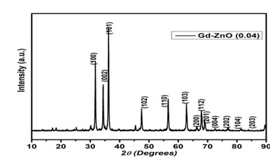
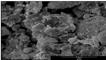
Figure 1 showed the Gd metal doped ZnO sample's XRD pattern. The JCPDS file #80-0075 and the crystal planes of the wurtzite ZnO structure balanced all of the diffraction peaks. The XRD patterns of synthetic samples of Gd substituted ZnO in the range of 2θ (diffraction angle) from 10° to 90° are displayed in 1, displaying all of the diffraction peaks [5]. Figure 1 displays the diffraction peaks that correspond to various hkl values, including (100), (002), (101), (102), (103), (200), (112), (201), and (202). Using FeSEM, figure 2 illustrates how the Gd ion affects ZnO shape when it is clicked out. The micrographs in the figure are polycrystalline in nature and exhibit agglomeration, asymmetric forms, and sizes as a result of the huge chemical reaction of energy.

jcm_37_1.jpg
Figure 1:
XRD Pattern of Gd doped ZnO nanostructures.

jcm_37_2.jpg
Figure 2:
FeSEM micrograph of Gd doped ZnO nanostructures

2. Photo-catalytic Test and Studies

jcm_37_3.jpg
Figure 3:
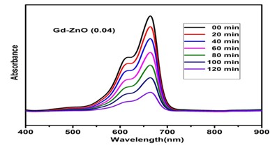
Photocatalytic Degradation of Gd doped ZnO nanoparticles.

jcm_37_4.jpg
Figure 4:
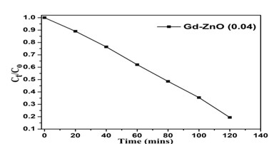
Variation of Degradation concentration ratio of Gd doped ZnO.


The obtained ZnO sample doped with Gd was investigated for photo-catalysis uses. The MB (methylene blue) and Malathion pesticide degradation studies were carried out using photo-catalysis in an aqueous solution with visible light present. In order to achieve a dark medium, the catalytic reactor assembly was placed in a cubical chamber for this study. A representative process involved placing 80 millilitres of dye solution (12.0 mg/Ltr) in a container with 1.10 g/Ltr of photo-catalyst. After 20 minutes of sonication, the powdered photocatalyst was evenly distributed throughout the dye solution. Because of the long chromophore, the distinctive absorption peaks of MB dye are situated at approximately 662 nm. The degradation of MB (organic pollutant) dye in the medium of a Gd-doped ZnO sample acting as a photo-catalyst is shown in Figure 3 with respect to time variations. Using the relation [6], the efficiencies of degradation (η) of the BCFO photocatalysts have been calculated:
jcm_37_5.jpg                   
where Ct is the absorbance intensity at time t = t and Cₒ is the initial absorbance intensity. Figure 4 displays the changes in the comparative rates (Ct/Cₒ) of the ZnO sample doped with Gd.

3. Photo-catalytic Mechanism

The presence of a possible catalyst, such as Gd-doped ZnO, is necessary to understand the process of photon-induced catalysis and to identify the precise mechanism involved in visible light-driven photocatalysis. Because of light absorption, light irradiation (hv ≥ Eg) on the photo-catalyst surface produces electrons-holes (e- - h+) in the band structure, which is crucial for the degradation of dyes or pesticides by generating hydroxyl radicals or free active radicals [7]. The hydroxyl or free radicals (O2-, -OH, etc.) that are generated during the glibness of e--h+ couples via H2O and O2 function as an active species in photon-induced catalysis.

Conclusions

In order to examine the impacts of Gd ions on the production process and its properties, the Gd doped ZnO sample was effectively prepared at 450 °C temperature using the co-perception technique. The XRD data supports the hexagonal structure of wurtzite. The crystallite sizes, planner distance,

and cell volume of Gd doped ZnO are expected to be 29 nm, 2.6243 Å, and 61.32 Å3, in that order. The FESEM images of the Gd-doped ZnO show the aggregation. For two hours, the photocatalytic degradation of Gd-doped ZnO nanoparticles was measured in the photocatalytic reactor. The Gd doped ZnO sample has 81% degradation efficiency (ɳ%), making it suitable for water purification in the dye industry.

Acknowledgment

The authors express their gratitude to Baba Mastnath University, Rohtak, for the facilities provided.

References

1. K. H. Kim, et al., “A comparative study on the structural properties of ZnO and Ni-doped ZnO nanostructures”, Materials Letters 149 (2015): 8-11.

2. A. Mclaren et al., “Shape and size effects of ZnO nanocrystals on photocatalytic activity”, Journal of the American Chemical Society 131.35 (2009): 12540-12541.

3. A. S. Menon, et al., “Studies on structural and optical properties of ZnO and Mn-doped ZnO nanopowders”, Indian Journal of NanoScience 1.1 (2013): 16-24.

4. B. Shohany et al., “Doped ZnO nanostructures with selected elements-Structural, morphology and optical properties: A review”, Ceramics International 46.5 (2020): 5507-5520.

5. J. Yang, et al., “Low-temperature growth and optical properties of Ce-doped ZnO nanorods”, Applied Surface Science 255.5 (2008): 2646-2650.

6. W. A. Sadik, et al., “The Use of heterogeneous advanced oxidation processes to degrade neutral Red Dye in aqueous solution”, Polymer-Plastics Technology and Engineering 43, no. 6 (2004): 1675-1686.

7. Y. H. Tan et al., “MoS2@ ZnO nano-heterojunctions with enhanced photocatalysis and field”,