E-ISSN:2583-9152

Research Article

Nano Materials

Journal of Condensed Matter

2023 Volume 1 Number 2 Jul-Dec
Publisherwww.thecmrs.in

A Comprehensive Examination of Ni@AgCl Nanoparticles – An Insight for Opto-Electronic Applications

Shivani RB1, R Vanathi Vijayalakshmi2*
DOI:10.61343/jcm.v1i02.29

1 Shivani RB, Department of Physics, Presidency College, Chennai- 600 005, Tamil Nadu, India.

2* R Vanathi Vijayalakshmi, Department Of Physics, Queen Marys College, Chennai600 005, Tamil Nadu, India.

In this study, chemical reduction approach was used to successfully manufacture Ni@AgCl core-shell nanoparticles with molar ratios of 1:1 and 4:1. The crystalline nature and face-centered cubic structure of these materials were confirmed by X-ray diffraction (XRD) investigation. Numerous characteristics, including crystallite size, dislocation density, lattice constants, volume of the unit cell, and lattice strain, were calculated using the Scherer formula and W-H plot. The findings showed that, in comparison to sample (B), which has a molar ratio of 4:1 sample (A) had a higher crystallite size and lower lattice strain. Both materials’ spherical shapes were validated by morphological analysis using EDAX, which also revealed their chemical composition. Indicating indirect band gap values of 2.53 eV and 2.45 eV, the UV-Vis absorption spectra of samples (A) and (B) revealed strong peaks at 237 nm and 239 nm, respectively, confirming their semiconducting characteristics. For samples (A) and (B), photoluminescence examination revealed emission wavelengths at 397 nm and 398 nm, respectively, falling in the UV zone. In light of these results, the synthesized Ni@AgCl core-shell nanoparticles are attractive candidates for Opto-electronic applications.

Keywords: XRD, W-H Plot, UV-Vis absorption spectra, Photoluminescence..

Corresponding Author How to Cite this Article To Browse
R Vanathi Vijayalakshmi, , Department Of Physics, Queen Marys College, Chennai600 005, Tamil Nadu, India.
Email:
Shivani RB, R Vanathi Vijayalakshmi, A Comprehensive Examination of Ni@AgCl Nanoparticles – An Insight for Opto-Electronic Applications. J.Con.Ma. 2023;1(2):156-159.
Available From
https://jcm.thecmrs.in/index.php/j/article/view/29/

Manuscript Received Review Round 1 Review Round 2 Review Round 3 Accepted
2023-11-10 2023-11-15 2023-11-20 2023-11-25 2023-12-01
Conflict of Interest Funding Ethical Approval Plagiarism X-checker Note
None Nil Yes 21.90%

© 2023by Shivani RB, R Vanathi Vijayalakshmiand Published by Condensed Matter Research Society. This is an Open Access article licensed under a Creative Commons Attribution 4.0 International License https://creativecommons.org/licenses/by/4.0/ unported [CC BY 4.0].

Introduction

By enabling unparalleled control over material properties at the nanoscale, nanotechnology has transformed a number of scientific disciplines [1]. In this setting, the intriguing class of materials known as Ni@AgCl core-shell nanoparticles has gained attention. These materials have potential uses in Opto-electronics, including those related to the military, telecommunications, automatic access control systems and acoustic devices [2]. Nickel has a wide range of uses in electronic devices and catalysis due to its magnetic characteristics and strong electrical conductivity [3]. On the other hand, silver is highly prized in optical systems and sensors due to its remarkable optical qualities, which also include high electrical conductivity and potent plasmonic effects. This core-shell nanoparticle is an appealing option for Opto-electronic applications due to the broad bandgap of silver chloride, which provides semiconductor-like properties to it [4]. This work aims to analyze the structural, morphological, and optical characteristics of core-shell nanoparticles made of nickel, silver, and silver chloride and to investigate their potential in opto-electronic devices. Additionally, modifying the nanoparticles for particular opto-electronic applications requires an understanding of the link between structural properties and optical responses [5].

Material Synthesis

Synthesis of Ni@AgCl Core-Shell Nanoparticles
In the synthesis process, 0.003M of Nickel chloride hexahydrate is dissolved in 50 ml of distilled water. 0.002M of sodium borohydride is dissolved in 50ml of distilled water. Then 10 ml of sodium borohydride is added drop by drop to the above solution, and stirred again for 24 hrs, continuously. Black colour precipitation ensures the formation of nickel nanoparticles. 0.001M of silver nitrate is dissolved in 50 ml of distilled water, and added to the above solution. Grey colour is precipitated, and the solution is allowed to stir 24 hrs continuously at room temperature. The solution is filtered and dried at 900 °C to get the fine Ni@AgCl nanoparticles. The same procedure is followed for molar ratio 4:1.

Results and Discussion

Figure 1(A) shows eleven distinct diffraction peaks

which were clearly observed at 2θ values of 28°, 32°4ˈ, 38°3ˈ, 44°5ˈ, 46°3ˈ, 54°9ˈ, 57°6ˈ, 64°5ˈ, 67°5ˈ, 74°5ˈ, 76°8ˈ. The major characteristic peak corresponds to the (111), (200), (220), (311), (222), (400), (331) crystal planes of the face centered cubic structure of AgCl with space group Fm3m and lattice parameters of  according to the JCPDS Ref. No: 851355. The crystal planes (111), (200), (222), (311) belong to the face centered cubic structure of Ag with space group Fm3m according to the JCPDS Ref. No: 893722 [6]. Figure 1(B) shows eleven distinct diffraction peaks at 2θ values 27°8ˈ, 32°2ˈ, 38°, 44°3ˈ, 46°1ˈ, 54°7ˈ, 57°4’, 64°3ˈ, 67°4ˈ, 74°4ˈ, 76°7ˈ which corresponds to the (111), (200), (220), (311), (222), (400), (331) crystal planes of AgCl. Similarly, the crystal planes (111), (200), (220), (311) belong to the Ag crystal structure. Finally, comparing the XRD patterns of the synthesized samples (molar ratio 1:1, 4:1), we identify that the major characteristic peak of the crystal plane (111) of sample (B) which belongs to the AgCl peak, has higher intensity than the sample (A). No typical diffraction peaks of Ni are observed, this may be due to the fact that Ni nanoparticles are covered by AgCl nanoparticles [7]. Table -1 display the structural parameters of Ni@AgCl nanoparticles of both molar ratios.

X-ray Diffraction Analysis

The XRD patterns of the synthesized Ni@AgCl core-shell nanoparticles of molar ratio 1:1 and 4:1 is reported in figure 1(A) and 1(B). From the XRD pattern, it is clear that majority of the peaks in samples (molar ratio 1:1, 4:1) are sharp indicating that it is highly crystalline in nature. Major peaks belong to AgCl and some of the peaks represent Ag.

jcm_29_1.jpg

Table 1: Structural parameters of Ni@AgCl core-shell nanoparticlesjcm_29_2.jpg


jcm_29_3.jpgFESEM Analysis

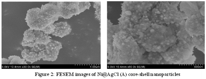
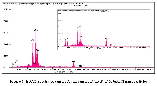
The surface morphology of the synthesized of Ni@AgCl core-shell nanoparticles of molar ratio 1:1 is reported in Fig 2. From the SEM images, we can find that Ni@AgCl core-shell nanoparticles are spherical in shape. The average sizes of the Ni@AgCl (A), Ni@AgCl (B) nanoparticles were calculated as 63.59 nm and 46.72 nm.jcm_29_4.jpgFigure 3 shows EDAX spectra of the synthesized Ni@AgCl  core shell nanoparticles of molar ratio 1:1 and 4:1. The elemental composition of the Ni@AgCl core-shell nanoparticles were confirmed by EDAX results. In this spectrum, it is seen that the major elements Ni and AgCl were present. Also, it is identified that the percentage of AgCl is higher than that of Ni element.
Optical Studies
UV-Visible Absorption Spectrum Analysisjcm_29_5.jpg
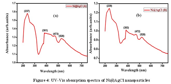
The UV-Vis absorption spectra of the synthesized Ni@AgCl core-shell nanoparticles of molar ratio 1:1 and 4:1 are reported in figure 4. The maximum absorption peak of the sample Ni@AgCl (A) was observed at 237 nm. Also, it gets three additional peaks at 381 nm, 473nm, and 528 nm. Reportedly, the highest absorption peak of 237 nm and 381 nm was assigned to AgCl.
From the absorption spectrum, the band gap energy was calculated by Tauc plot. The band gap energy was found to be an indirect band gap and the value is 2.53 eV. Similarly, the maximum absorption peak of the sample Ni@AgCl (B) was observed at 239 nm and the major peak is assigned to AgCl. The band gap energy was found to be an indirect band gap and the value is 2.45 eV. Though, the indirect band gap of AgCl value is 3.28 eV by reference, [8] when it is coated by the nickel element, the band gap reduces to 2.53 eV and 2.45 eV for the sample A and B respectively. From this result, we infer that the synthesized sample may be a semiconducting material [9].

Photoluminescence Analysis

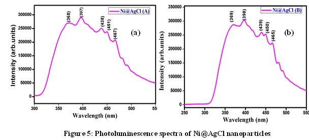
Figure 5 shows the PL spectra of synthesized Ni@AgCl core-shell nanoparticles of molar ratio 1:1 and 4:1 [10]. The emission wavelength of synthesised Ni@AgCl (A) core-shell nanoparticles of molar ratio 1:1 was identified at 397 nm and sub bands on both sides of the major peak was observed. The emission wavelength of synthesized Ni@AgCl (B) core shell nanoparticles of molar ratio 4:1 was identified at 398 nm [11,12].jcm_29_6.jpgFrom the PL spectra of the samples A and B, it is clear that the emission of photons is observed at UV region [13]. The sub bands that are responsible for the surface states, play key role in detecting oxygen vacancies and defects on the surface. The synthesized samples Ni@AgCl (A) and Ni@AgCl (B) acting as a semiconducting material and emitting photons at UV region can be used for acoustic devices and optoelectronic applications [14].

Conclusion

Ni@AgCl core-shell nanoparticles of molar ratio 1:1 and 4:1 have been successfully synthesized by chemical reduction method. The XRD results indicate that the synthesized samples are crystalline in nature. The SEM images of the synthesized sample indicate that both the samples were spherical in shape. The UV-Vis absorption


spectrum of the samples A and B indicates sharp absorption peak at 237 nm and 239 nm respectively and the indirect band gap energy was found to be 2.53 eV and 2.45 eV respectively. From the photoluminescence analysis, the emission wavelength is observed at 397 nm and 398 nm respectively for the samples A and B. The emission of light of both the samples falls on the ultraviolet region. From the above results, we infer that the synthesized sample Ni@AgCl (A) and Ni@AgCl (B) which gets excited at 237 nm and 239 nm emits light at UV-region can be used in Opto-electronic applications such as in military services, telecommunications, automatic access control system, medical equipment and acoustic devices.

References

1. Zhou Y, Zhao L et al., J Alloys Compd. 2023, 935, 168012.

2. Zhang C, Chen J et al., Adv Funct Mater. 2021, 31, 2100438.

3. Seong C, Ryu H, et al., Catal Sci Technol. 2023.

4. Alruqi M, Rabia M et al., J Appl Polym Sci. 2023, 140, 19.

5. Abomostafa HM., J Inorg Organomet Polym Mater. 2021, 31, 2914.

6. Saravanan R, Khan MM, RSC Adv. 2015, 5, 346451.

7. Han SK, Gu C et al., J Am Chem Soc. 2015, 137, 5396.

8. Cui B, Wang C et al., J Colloid Interface Sci. 2020, 578, 10.

9. Kim HR, Kim YD et al., Sens Actuators B Chem. 2004, 97, 354.

10. Xiong ZW, Cao LH et al., Appl Surf Sci. 2018, 445, 65.

11. Aarjane M, Slassi S et al., J Mol Struct. 2020, 1199, 126990.

12. Zheng Z, Wang T et al., J Colloid Interface Sci. 2022, 606, 47.

13. Xu G, Xu Z et al., Chem Sus Chem. 2017, 10, 2903.

14. Zhang G, Li G et al., Chem Cat Chem. 2015, 7, 2870.