E-ISSN:2583-9152

Research Article

Nano Materials

Journal of Condensed Matter

2024 Volume 2 Number 1 Jan-Jun
Publisherwww.thecmrs.in

Effect of Cation Disorder on Basic Parameters of ZnFe2O4 Nano-Particles Synthesized by Honey Mediated Solution Combustion Technique

Raghuvanshi S1*
DOI:10.61343/jcm.v2i01.39

1* Saroj Raghuvanshi, Shri Cloth Market Institute Of Professional Studies, Indore, Mp, India.

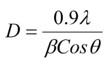
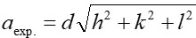
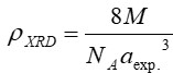
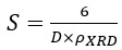
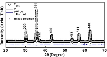
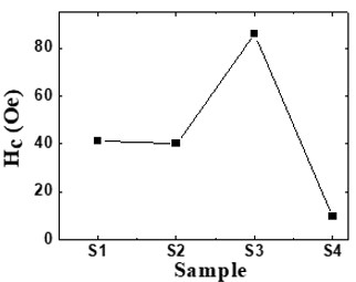
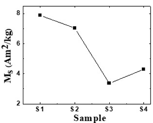
Nanotechnology contracts by the invention and practice of material using nanoscale dimension. Nanoscale measurement delivers nano-particles a bulky superficial area ‘S’ to volume ratio, hence very specific characteristics. Bulk zinc ferrite (ZnFe2O4) exhibits anti-ferromagnetism, with Néel temperature of 10K, is paramagnetic at room temperature. It has a typical spinel structure and exclusive tetrahedral - A site preference when Zn2+ is present, whereas Fe3+ ions occupy the octahedral - B site. Cationic disorder induced fractional overturn of the spinel structure, owing to partial immigration of Fe3+ ions from B to A site can prompt ferrimagnetism in nano zinc ferrite. Owing to the high concentration of hazardous substances and harsh conditions involved in the chemical and physical manufacturing process, a green approach utilizing fungi, bacteria, and plants has been used. Present work reports comprehensive study of the synthesis, structural and magnetic investigation of room temperature ferrimagnetism in ZnFe2O4 nanoparticles, prepared by sol gel auto-combustion mode and green synthesis method. Effect of conventional thermal annealing (ann. at 600oC for 3 hours) on magnetic properties is also reported. The magnetic and structural characteristics of synthesized and annealed ZnFe2O4 samples were determined by vibrating sample magnetometer (VSM) and X-ray diffraction (XRD). XRD verifies that the samples have formed a single-phase nano-crystalline cubic spinel configuration.

Keywords: Zn Ferrite, Green Synthesis, Cationic Disorder

Corresponding Author How to Cite this Article To Browse
Saroj Raghuvanshi, , , Shri Cloth Market Institute Of Professional Studies, Indore, Mp, India.
Email:
Raghuvanshi S, Effect of Cation Disorder on Basic Parameters of ZnFe2O4 Nano-Particles Synthesized by Honey Mediated Solution Combustion Technique. J.Con.Ma. 2024;2(1):1-5.
Available From
https://jcm.thecmrs.in/index.php/j/article/view/39

Manuscript Received Review Round 1 Review Round 2 Review Round 3 Accepted
2023-12-04 2023-12-12 2023-12-20 2023-12-28 2024-01-04
Conflict of Interest Funding Ethical Approval Plagiarism X-checker Note
None Nil Yes 19.63

© 2024by Raghuvanshi Sand Published by Condensed Matter Research Society. This is an Open Access article licensed under a Creative Commons Attribution 4.0 International License https://creativecommons.org/licenses/by/4.0/ unported [CC BY 4.0].

Introduction

Nano ferrites are being comprehensively investigated owing to their potential applications in magnetic fluids, drug delivery, magnetic refrigeration, high-density information storage, etc. [1]. Spinel ferrites have been intensively examined due to their multipurpose chemical and physical properties, due to their scientific and technical applications in photocatalysts, magnetic sensors, nano-electronics, bio-sensors, bio-medical [2]. These applications are based on the basic construction and magnetic performance of spinel ferrites which can be tailored through selecting proper preparation method. Multiple kinds of synthesis approaches including hydrothermal, sol gel, co-precipitation, electrochemical, and others have been applied for preparation of ferrite nano-particles. Cations are circulated between tetrahedral ‘A’, and octahedral ‘B’ sites in spinel ferrites, where the structural and magnetic atmospheres are rather dissimilar. Consequently, the information of the cation arrangements (on site ‘A’, ‘B’) is essential to recognize magnetic behavior of spinel ferrites. Cation distribution can be different: i) if the sample is synthesized by a particular method (owing to dissimilar preparation conditions in each method), ii) substitution by particular ions and iii) post preparation thermal treatments which can be profitably utilized to have control on structure, this will therefore impact the ferrites’ magnetic characteristics. Thermal annealing would lead to changes in crystallite size as well as re-distribution of cations, leading to changes in structural and magnetic properties.

Latest developments in nano-technology are prominence on nature friendly, cost-efficient preparation methods. The green approach to nanoparticle synthesis is a non-toxic, environmentally beneficial way to synthesize nanomaterials from natural resources. Safe nanotechnology is entering a new phase because to this environmentally friendly approach. Natural honey is known as the world’s oldest food source with high energy and nutritious value [3] Honey is sweet (viscous) fluid made by bees and the major residents of honey are fructose and glucose [4]. Regular honey contains glucose and fructose, which contribute to the creation of nanoparticles that act as a natural reductant, sticky medium, and protective mediator. By using honey to prepare

spinal ferrites, harmful and toxic reducing mediators are avoided during the combustion process. Special chemical properties of honey extract are used in environmentally friendly nanoparticle production. Method mediated by honey, offers a number of benefits over the traditional method that uses citric acid as a mediator.

Zinc ferrite (ZnFe2O4) is one of the most significan­t industrial substantial, which finds use in radio engineering, semiconductors, radio technology, and other fields. The magnetic properties of Zn ferrite nanoparticles were observed to have higher magnetization values related to the bulk materials and also the synthesis technique might also play imperative role in achieving the ferromagnetic nature of Zn ferrite [5]. Several reports on ZnFe2O4 have showed that, ZnFe2O4 is antiferromagnetic at room temperature with Néel temperature TN ≈ 10 K [6]. But, observed ferromagnetic nature of ZnFe2O4 synthesized by various techniques shows creation of oxygen positions in the structure. Bulk zinc ferrite ha­­­ve the normal spinel structure with Zn2+ ions occupying tetrahedral (A) site and Fe3+ ions being occupied in octahedral (B) site. [7]. The magnetic properties of Zn ferrites significantly depend when a small amount of Fe3+ ions migrate towards A site [8], therefore non-equilibrium cation distributions become a tool for tailoring magnetic properties. In this work, ZnFe2O4 spinel ferrite was synthesized using citric acid and a natural substance called honey. Further, the effects of citric acid, natural products on cation distribution, structural changes, and magnetic parameters were examined.

Sample Synthesis and Characterization 

ZnFe2O4 nanopowders were prepared by a solution ignition method with acetate / citrate–nitrate pre-cursors and honey. The prepared mixture was heated to 120 degree Celsius in the air to produce "dry gel," or loose powder. The internal-combustion reaction is a heat-releasing oxidoreduction process in which nitrates mixture and honey act similarly to conventional oxidants and fuels. Samples that were prepared were annealed at 600 oC for 3hrs to expand the degree of crystallization. Dry gel samples using critic acid and honey as fuels were labelled: citric acid as S1, honey as S2 while heat treated samples were categorized as: citric acid as S3, honey as S4. The magnetic and structural features of synthesized and annealed ZnFe2O4


samples were determined by vibrating sample magnetometer (VSM), and X-ray diffraction (XRD). Reitveld alteration software ‘MAUD’ (Material Analysis Using Diffraction) [9] was used for complete proof study of XRD patterns to calculate experimental lattice parameter (aexp.). Distribution of Cation among varies sites was determined by XRD intensities, using Bertaut method, as described in [10]. Using the obtained cation distribution ionic radii of A-site (rA) and B-site (rB) (The radius of the cations are taken from the work of Shannon) [11], theoretical lattice parameter (ath.), Oxygen positional parameter (u43m at A-site) and (u3m at B-site), the bond angles (θ1, θ2, θ3, θ4, θ5), and surface area (S) were calculated as described in [12 -14].

Grain size is obtained by strongest diffraction peak [311], by using Scherrer’s formula,

jcm_39_1.jpg

Where β - Line width, λ - Wavelength of x-ray used, θ - Peak position (in 2θ scale).

For plane [311], the Lattice parameter (aexp) was computed using the formula,

jcm_39_2.jpg

Where d - Inter planner distance, (h, k, l) - Miller indices.

Following equation was used to compute the X-ray density (ρxrd),

jcm_39_3.jpg

Where NA – Avagrodo’s Number, M – Molecular weight, aexp – Lattice parameter.

Calculation of Specific surface area (S) was done by using formula,

jcm_39_4.jpg

Where D - grain diameter, rXRD - x-ray density.

The hopping distance for site A and B (LA and LB)

jcm_39_5.jpg

           Where aexp – Lattice constant.

Following formula was used to get cation distribution,

jcm_39_6.jpg

Where  Iobshkl- experimental and Icalhkl  - calculated intensities for reflection (hkl).

Results and Discussion

The representative rietveld refined XRD plot shown in figure 1 for the selected ZnFe2O4 composition (S3) of the annealed sample. XRD patterns for prepared S1, S2 and annealed S3, S4 samples support single phase cubic spinel structure formation, ascribed honey and citric acid both plays the role of good chelating agent. Annealed (600 oC for 3hrs.) ZnFe2O4 samples support single phase formation (secondary phase not present) of nanodimensional cubic spinel (Fd3m space group) structure, attributed as role of thermal treatment support the development of spinel phase. The structural factors: Scherrer’s grain dimension (D), x-ray density (ρXRD), and specific surface area (S) of as prepared and heat-treated samples calculated by examining XRD data, are showed in table-1. Grain size of as prepared samples (S1, S2) range between 22.84 – 32.52 nm, where grain size of annealed samples (S3, S4) range between 53.37 – 61.71 nm evidently illustrates formation of nano-dimensional spinel ferrite. Grain size of annealed samples (S3, S4) rises as compared to as-prepared (S1, S2) samples is ascribed to result of heat treatment that supports grain growth of the samples. Variation in D of the studied samples with citric acid and honey is attributed to the effect of reaction condition, which favors the formation of new nuclei preventing further growth of particles [15]. Figure 2 depicts the change of experimental (using XRD data), theoretical lattice space (aexp., ath.) of dry gel and annealed samples. The observed changes in aexp., ath of samples S1 – S4 can be ascribed to the performance of fuel (citric acid and honey), which help to relocation of Zn2+ and Fe3+ ions having dissimilar ionic radius, which alter the microstructure of sample. The excellent surface-to-bulk ratio of nano ferrites makes them a vital component in improving the performance of solid catalysts. Specific surface area (S) is an inversely correlated with both Scherrer’s grain diameter (D)


and x-ray density (ρXRD). Particles specific surface area is the summation of the areas of the exposed surfaces of the particles per unit mass. [16]. Specific surface area of the studied samples (S1, S2, S3, S4) varies from 18.15 - 48.64 m2/g. For sample S1, the high specific surface area value and low D value obtained make it a better option for the purpose of catalysis.

XRD analysis was used to determine the cation dispersal, which is shown for each sample in the second table. It is noteworthy fact, Zn ferrite displays normal spinel structure. The Zn2+ and Fe3+ ions' occupation of sites A, and B provides a clear picture of the cation distribution of the materials under study. According to the closely matched aexp., ath, the estimated and real distribution of cations between the sites A and B agree extremely well. In the present work, the non-magnetic Zn2+ ions (zero magnetic moment) are dispersed at both A, B sites which is responsible for relocation of magnetic Fe3+ ions reside on site B. A precise depiction of the function of the natural extract and citric acid is reflected in obtained cationic distribution.

Table 1: X-ray density (rXRD), surface area (S), Grain diameter (D) of Zn ferrite.

SampleD(nm)rXRD(Kg/m3)S(m2/g)
S122.845400.148.64
S232.525365.234.39
S353.375350.721.01
S461.715357.118.15

jcm_39_7.jpg
Figure 1: XRD pattern of nano sized zinc ferrite

jcm_39_8.jpg
Figure 2: Variation of aexp and ath

Observed results shows noticeable variation in XRD and cation distribution parameters like Oxygen positional parameter (u43m at A-site) and (u3m at B-site), ionic radii of A-site (rA) and B-site (rB), hopping length for A-site (LA) and B-site (LB), (see table 2), bond positions (θ1, θ2, θ3, θ4, θ5) (see table 3). Oxygen position parameter u43m for tetrahedral site range between 0.3816 - 0.3857 and u3m at B-site is range between 0.2566 - 0.2607 (shown in table 2). It ought to be pointed out that u43m and u3m is a measurement for the sample’s disorganisation, and its standard value of u43m is 0.375 (not any disarray in the lattice). Experimental values of oxygen position parameter for all calculated specimens are in excess of the real value indicating that the spinel's tetrahedral and octahedral sites are distorted. Variation of rA, and rB correspondingly spans between 0.054 nm – 0.60 nm, and 0.65 nm – 0.68 nm. Observed changes in rA and rB values of studied samples show the variation of distance between tetrahedral and octahedral cation-anion.

Table 2: Cation distribution, changes in ionic radii of A-site (ra) and B-site (rb), hopping length for site A and B (LA and LB), Oxygen positional parameter (u43m at site -A) and (u3m at site- B), of ZnFe2O4 system.            

Cation Distributionsra (nm)rb(nm)U43mU3mLa(nm)Lb(nm)
S1(Zn0.41Fe0.59)A[Zn0.59Fe1.41]B0.0540.0670.38160.25660.36380.2970
S2(Zn0.55Fe0.45)A[Zn0.45Fe1.55]B0.0560.0680.38250.25780.36420.2973
S3 (Zn1.0)A[Fe2.0]B0.0600.0650.38570.26070.36490.2983
S4 (Zn0.9 Fe0.1)A[ Zn0.1Fe1.9]B0.0590.0650.38500.25990.36470.2978

Hoping length for sites A and B (LA, LB) are depicted in table 2, Changes that were noticed in hopping length for site A and B of studied specimens is similar to that of the variation of experimental lattice parameter.

The total strength of the magnetic exchanges (A-B, B-B, and A-A) is determined by the bond length and bond angles between the cations and cation-anion. Bond length is inversely correlated with strength (exchange cooperation), but directly connected to bond angle. Changes in bond angles among A-O-B (θ1, θ2), B-O-B site (θ3, θ4) and A-O-A site (θ5) in the examined specimens is illustrate in table 3, suggests about the magnetic interaction among A and B sites cations. Obtained highest value of bond angles θ1, θ2, θ5 and lowest value of bond angles θ3, θ4 for S1 indicates weakening


of the B-B relationship and rising of A-B, A-A interaction in comparison of the other samples.

jcm_39_10.jpg

jcm_39_11.jpg
Figure 3: (a) Variation of coercivity (Hc), (b) Variation of saturation magnetization (Ms)

Figure 3 (a) and 3 (b) respectively depicts the variation of coercivity (Hc) and saturation magnetization (Ms) of the examined specimens. Coercivity values of samples range between 09.54 – 86.00 Oe. Perusal of Fig. 3(a) shows that highest value of Hc for sample S3 suggests effect of heat treatment. Figure 3 (b) depicts variation of experimental Ms (at 300 K) of studied samples. Highest Ms of 7.89 Am2/kg was calculated for the as-burnt S1 sample in contrast to other samples, whereas annealed sample S3 gives value of Ms is 3.37 A m2/kg. It should be mentioned that the results obtained with honey and citric acid are a result of the Fe and Zn ions’ varying friction on the sites A, B. (table 2). Obtained changes in cation distribution (on A, B site) increases alteration (u) within the spinel system which is amply demonstrated by detected deviations in Ms

, shows the significance of cationic dispersion in influencing determining magnetic characteristics.

Table 3: Changes in bond angles ((A-O-B) θ1, θ2, (B-O-B) θ3, θ4, (A-O-A) θ5) of ZnFe2O4 system.

Sampleθ1 (o) (AOB)θ2(o) (AOB)θ3 (o)(BOB)θ4 (o) (B-O-B)θ5 (o) (A-O-A)
S1123.142144.10793.155125.98173.961
S2123.082142.71693.860125.35273.190
 S3121.865138.86595.218126.42970.635
S4122.082139.71694.860126.35271.190

Conclusion

In summary, ZnFe2O4 nanoparticles are made by using a simple sol-gel auto combustion process that uses honey and citric acid as combustibility. The structural phase development of ZnFe2O4 spinel ferrite nanoparticles was demonstrated by the x-ray diffraction investigation. Cation distribution confirmed the migration of Zn element, Fe ions in mutually tetrahedral and octahedral sites in synthesized ZnFe2O4. Grain size, structural alterations, and cation distribution are observed to have a notable effect on the magnetic properties of zinc ferrite nanoparticles. The cation dispersal and grain size effect were discovered to cause changes in magnetic characteristics such as coercivity and saturation magnetization.

Acknowledgement

I would like to acknowledge my guide Prof (Dr.) S. N. Kane, Magnetic Materials Laboratory, SOP, DAVV, Indore, with my sincerest respect and gratitude, who not only introduced me to this area of Nanocrystalline Ferrites but also explained the finer aspects with a lot of patience and determination throughout.

References

1. D Peddis, N Yaacoub, M Ferretti, A Martinelli, G Piccaluga, A Musinu, C Cannas, G Navarra, J M Greneche, D Fiorani, Phys.: Cond. Matter, 23:426004, 2011.

2. H Agarwal, S Venkat Kumar, and S Rajeshkumar, Resource Efficient Technologies, 3: 406–413.2017.

3. D Philip, Spectrochim. Acta A Mol. Biomol. Spectrosc., 73(4):650–653, 2009.

4. E R Balasooriya, C D Jayasinghe, U A Jayawardena, R W D Ruwanthika, R M


de Silva, and P V Udagama, Journal of Nanomaterials, Vol. 2017:5919836, 2017.

5. C E R Torres, F Golmar, M Ziese, P Esquinazi, and S P Heluani, Phy. Rev. B, 84:064404, 2011.

6. L Raghavan, G Pookat, H Thomas, S Ojha, D K Avasthi, and M R Anantharaman, J. Magn. Magn Mater., 385:265–271, 2015.

7. H Ohnishi, and H T Teranishi, J. Phys. Soc. Jpn. 16:3543, 1961.

8. L Weil, E F Bertaut, and L Bochirol, J. Phys. Radium, 11:208, 1950.

9. L Lutterotti and P Scardi, J. Appl. Cryst., 23(4):246252, 1990.

10. L Gastaldi, and A Lapiccirella, J. Sol. Stat. Cham., 30:223229, 1979.

11. R D Shannon, Acta Cryst., A32:751, 1976.

12. J Smit, and H P J Wijn, Ferrites, New York: Wiley-Inter Science 1959.

13. K E Sickafus, J M Wills, and N W Grimes, J. Am. Ceram. Soc., 82:32793292, 1999.

14. X Qi, J Zhou, Z Yue, Z Gui, and L Li, Mater. Sc. and Eng. B, 99:278281, 2003.

15. R S Yadav, I Kuritka, J Vilcakova, J Havlica, J Masilko, L Kalina, J Tkacz, J Švec, V Enev, and M Hajdúchov, Adv. Nat. Sci.: Nanosci. Nanotechnol, 8:045002 (14pp), 2017.

16. Z X Tang, C M Sorensen, K J Klabunde and G C Hadjipanayis, Phy. Rev. Letters, 67:36023605, 1991.